3D OCT eye scan is an advanced method of detecting and monitoring the development of eye conditions including age-related macula degeneration (AMD), glaucoma, diabetes and retinal detachment.

3D OCT eye scanallows measurement of corneal shape (topography) and thickness for patients who wish to have laser refractive surgery.

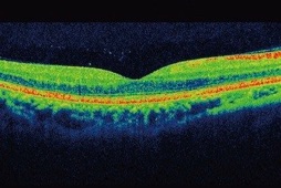
OCT Observed Conditions

'Dry' macular degeneration

This type of AMD affects 80% of people with macula degeneration. There is currently no treatment for this condition although vitamin supplements may slow down its progression.

'Wet' macular degeneration

This type of AMD often develops quickly in a matter of a few weeks. Early OCT scanning and referral allows treatment to be undertaken with anti-VEGF therapy drugs such as Lucentis which is injected into the eye.
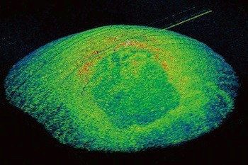
Glaucoma

Glaucoma damages the nerve fibres of the  optic nerve leading to blind spots in the vision. 3D OCT eye scan detects the damage caused by glaucoma by measuring the nerve fibre layers in the retina.

DIabetic Retinopathy

All stages of diabetic retinopathy can be detected with 3D OCT eye scan including the earliest stage involving macula oedema.

Floaters

Floaters can often be detected by the 3D OCT eye scan. Sometimes floaters are associated with ‘flashes’ especially if they are caused by posterior vitreous detachment or retinal detachment.

Retinal detachment

The OCT B scan shows a horizontal split in the retinal layers indicating retinal detachment. OCT allows highly accurate assessment of the extent of the condition for surgical purposes                   

OCT Your Questions Answered

OCT is analogous to an ultrasound scan for the eyes. It uses near-infrared light to measure the thickness of the different layers that make up the retina at the back of the eye.

Ideally, everyone should have a 3D OCT eye scan to ensure their eyes are healthy. 3D OCT is able to detect developing eye abnormalities which often start long before you notice your sight being affected. For those with existing eye conditions such as Age-Related Macular Degeneration (AMD), diabetic eye disease or retinopathy and glaucoma, 3D OCT should be performed at regular intervals to determine if the condition is getting worse. For example, 10-15% of ‘dry’ macula degeneration cases develop into the more serious ‘wet’ form. In a very short time, major changes in the eye can lead to severe and permanent loss of vision. The cutting-edge 3D OCT comparison software can detect the slightest deterioration in a progressing eye condition often allowing prompt remedial treatment.

If your eyes are healthy you should have a 3D OCT eye scan every time you have a routine eye examination. This will usually mean every 1 or 2 years.

Regular screening 3D OCT eye scans are essential if we are to detect eye disease at an early stage and before vision is permanently damaged. Many eye conditions, including diabetic retinopathy, progress very slowly and symptoms do not appear until it is too late for any treatment to be effective. If you suffer from a specific eye disease your optometrist will be pleased to tell you how often you should have a 3D OCT scan.

The fee for performing a screening 3D OCT eye scan is £40.00. If this initial scan reveals an abnormality of your eyes additional scans may need to be performed before we are able to refer you for further ophthalmological investigation (either via the NHS or privately). Performing the extra scans, where necessary, analysis of the results, report writing and liaising with your GP and eye consultant will incur an additional cost of £50.00.
Our 3D OCT eye scan service is not considered to be part of a normal NHS eye examination and therefore has to be paid for privately by NHS patients. Patients will be asked to pay for the 3D OCT eye scan in addition to any normal eye examination fees.

Our optometrist will advise you if there are any serious concerns about your eyesight and will arrange to send a written report to your GP. Your GP will, in turn, then arrange for you to be seen by a consultant ophthalmologist, usually at a local hospital.  If you have to return to us for further scans then comparative data will also be able to be collected to indicate any worsening of your eye condition. This information will ensure rapid, accurate diagnosis and can help an ophthalmologist decide on the optimum method of treating your condition, thereby improving the chances of you having a good visual outcome.

Yes, although the 3D OCT uses laser light to measure your eyes no harm can be done to any eye tissue irrespective of how many times the scan is repeated.

Normally no drops are required to enlarge the pupils of your eyes when a 3D OCT eye scan is made. Occasionally some people have very small pupils which prevent satisfactory measurements being taken . If this is the case for you, we will advise you and arrange for pupil dilation to be undertaken prior to the scan being performed. This may involve you revisiting our practice on an alternative date and a small additional charge may be made.

No, the OCT instrument does not make contact with your eyes. There is a short flash of light which occurs during the scanning process, but this is not uncomfortable.

Most of the eye conditions a 3D OCT eye scan can help diagnose are treatable providing they are detected at an early enough stage. Glaucoma, diabetic retinopathy and ‘wet’  AMD are some of the commonest conditions which can potentially cause blindness. Now that their effects on the eyes can be much more closely followed with 3D OCT eye scans we are often able to save sight that, just a few years ago, would have been permanently lost.